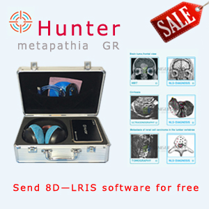
Bioplasm nls-research application area and improvement of images quality
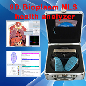
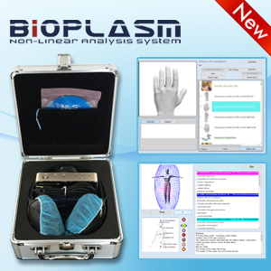
Technological innovations of the recent years in equipment for non-linear diagnostics and software resulted in cardinal extension of Bioplasm nls-research application area and improvement of images quality. One of the promising directions for the development of Bioplasm nls-diagnostics is a mode of multidimensional virtual reconstruction of an Bioplasm nls-image, allowing to visualize crosscuts and anatomical reconstruction of a researched area, which remain invisible during application of two-dimensional scanning only. This method offers the possibility of data manipulating, using rotation and zooming, rendering true anatomy of organs in a direct projection. Three-dimensional Bioplasm nls-scanning has a certain technical and practical advantages in visualization of anatomical and pathological structures in comparison with two-dimensional visualization. The method allows to make a crosscuts of acquired image, for example frontal ones, or to isolate separate areas in Fast Vision mode. It makes possible to see an internal structure of a parenchymatous organ, and if there are areas of affection inside the investigated region, an operator can isolate and zoom in areas of a parenchyma and thus see intratissual growths – in Bioplasm nls-ultramicroscanning mode. Spectral-entropic analysis (SEA) mode allows to define morphological character of a researched area by comparison of spectral similarity with etalon processes.

This article is provide from [Bioplasm nls],please indicate the source address reprinted:http://www.bioplasm-nls.com/nls_knowledge/Bioplasm_nls_research_application_area_and_improvement_of_images_quality.html