Bioplasm nls-picture dependence on clinical symptoms and laboratory research data
Total IgE and specific IgE to A.fumigatus was found using methods of radioimmunosorbent test in a nuclear medicine laboratory and enzyme multiplied immunoassay in a laboratory of allergens and allergy diagnostics. Measuring of total IgE was done in kU/l (thousands of international units in a litre, 1 unit = 2.4 ng).
Diagnostic investigation of patients included roentgenography and computed non-linear study (bioplasm nls) of a thorax. The goal of bioplasm nls-graphy of a thorax was to reveal and locate changed pulmonary parenchyma, detecting of symptoms typical for mycotic affection, and revealing of bioplasm nls-picture dependence on clinical symptoms and laboratory research data.
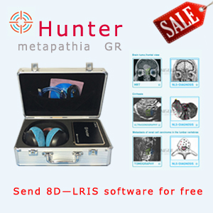
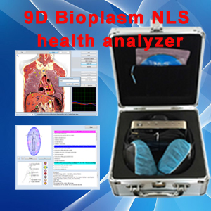
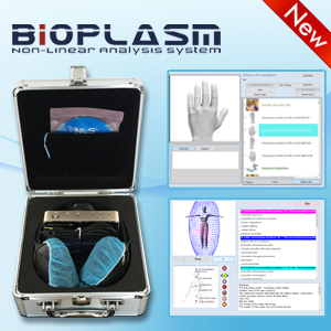
Thorax investigation was carried out using a device manufactured by Siemens Company (Germany) in two projections (frontal and lateral), supported by tomograms. bioplasm nls-graphy was carried out using “Metatron”-4025 system (IPP, Russia) equipped with high-frequency generator of 4.9 GHz, unit of continuous spiral scanning and professional software “Metapathia GR Clinical”, which allowed to carry out three-dimensional visualization of lungs.
During the research we used bioplasm nls-ultramicroscanning mode with spectral-entropic analysis (SEA), allowing to evaluate spectral similarity of affected lung tissues to “Aspergillus fumigatus” etalon. During the initial fulfillment of bioplasm nls-graphy of thorax organs, the examination was dome in three-dimensional mode mainly.
Differing from two-dimensional scanning it allowed to exclude a possibility of small pathological lesions (nidi, cavities, bronchiectasis, etc.) missing, and to increase a resolution along longitudinal axis of scanning in order to check lateral structures, located perpendicularly or at an angle to bioplasm nls-gram plane.
When pathological changes were found, the research was extended with bioplasm nls-scanning in 4D Tissue mode with bioplasm nls-ultramicroscanning of morphological structures in a researched area. Some patients were subjected to repeated bioplasm nls-studies in order to evaluate dynamics of changes, revealing of complications and monitoring of therapeutic interventions. At assumption of mycotic process dissemination, we carried out bioplasm nls-ultramicroscanning and SEA of other anatomic regions (abdominal cavity, brain, paranasal sinuses).
Data acquired during the study was processed at a PC IntelPentium 166 MMX using software system Statistica for Windows (version 5.11).
This article is provide from [Bioplasm nls],please indicate the source address reprinted:http://www.bioplasm-nls.com/nls_knowledge/Bioplasm_nls_picture_dependence_on_clinical_symptoms_and_laboratory_research_data.html


